ULTRA (samfilcon A) Contact Lenses
Bausch + Lomb
INDICATION FOR USE:
Single Vision Spherical (SVS) Vision Correction : For extended wear for up to 7 days; correction of refractive ametropia (myopia and hyperopia) in aphakic and/or not-aphakic persons with non-diseased eyes, exhibiting astigmatism of 2.00 diopters or less, that does not interfere with visual acuity.
Presbyopia Vision Correction: For extended wear for up to 7 days; correction of refractive ametropia (myopia, hyperopia and astigmatism) and presbyopia in aphakic and/or not-aphakic persons with non-diseased eyes, exhibiting astigmatism of 2.00 diopters or less, that does not interfere with visual acuity.
Astigmatism Vision Correction: For extended wear for up to 7 days; correction of refractive ametropia (myopia, hyperopia and astigmatism) in aphakic and/or not-aphakic persons with non-diseased eyes, exhibiting astigmatism up to 5.00 diopters.
DEVICE DESCRIPTION:
- Contact Lenses are 46% water and 54% samfilcon A material
- Samfilcon A material is a hydrophilic copolymer of siloxane methacrylate, a siloxane cross-linker, and N-vinyl pyrrolidone
- Tinted blue for visibility with Reactive Blue Dye 246, a color additive that conforms to 21 CFR Part 73.3106
- Utilizes MoistureSeal® technology
- In its hydrated state, when placed on the cornea acts as a refracting medium to focus light rays on the retina
EFFECTIVENESS:
- Prospective, multi-center, two-arm cohort study, randomized, double-blinded, 12-month clinical study, n= 816, B+L ULTRA vs. B+L PureVision control group
- Primary effectiveness endpoint: High contrast, distance visual acuity with dispensed lenses at the 12-Month Follow-up Visit. 97% of subject eyes achieved at least 20/25 with ULTRA
- Secondary effectiveness endpoint: Lens wear time reported as average extended lens wear time (days/week) since last visit. Average wearing time was 6.7 (±0.031) days for both groups
- Line change in visual acuity from baseline: 3.0% eyes in Ultra group vs. 3.8% eyes in PureVision group experienced worsening of ≥ 2 lines (≥ 10 letters)
- Unfavorable lens performance: Higher proportion for PureVision group
SAFETY:
- The primary safety endpoint: Rate of serious or significant non-serious adverse
events during the 12-month follow-up: No serious AEs for either lens group - 3.0% of Ultra eyes experienced significant non-serious AEs vs. 2.4% of PureVision eyes – Noninferiority was met using a predetermined threshold of 5.0%.
REGULATORY PATHWAY: PMA
- Device Procode: LPM

ThinPrep Integrated Imager
Hologic, Inc.
INDICATION FOR USE: Uses computer imaging technology to assist in primary cervical cancer screening of ThinPrep® Pap Test slides for the presence of atypical cells, cervical neoplasia, including its precursor lesions (Low Grade Squamous Intraepithelial Lesions, High Grade Squamous Intraepithelial Lesions), and carcinoma as well as all other cytologic criteria as defined by the Bethesda System: Terminology for Reporting Results of Cervical Cytology
DEVICE DESCRIPTION:
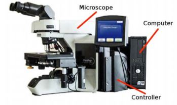
Three major subsystems
- Microscope: Imaging camera, slide ID reader, automated stage, hand controls and adjustable touch screen user interface
- Controller: Controls the electromechnical components of the Microscope
- Computer: Hosts system application software and system database
Two major functions
- Imaging: Takes high magnification frames at more than 500 x-y locations covering entire cell spot. z-locations (focal plane) calculated based on x-y locations. Software
analyzes images of cell spot, identifies objects of interest based on optical density - Review: Retrieves locations of the objects of interest and sequentially positions for evaluation and interpretation by the cytotechnologist (CT)
Two work modalities
- Sequential: Slide is imaged and then reviewed immediately by the CT.
- Batched: Slides can be imaged in succession, with the coordinates stored in the
computer database, for review by the CT or pathologist at a later time.
EFFECTIVENESS:
- Multi-center, two-armed clinical study, n= 1,260 patient cases that covered all cytologic diagnosis categories, similarity of ThinPrep Integrated Imager (TI) to ThinPrep Imaging System (TIS)
- Significantly higher sensitivity and slight decrease in specificity, with TI
- Workload assessment: The number of slides that a CT can scan and review in one day is less on I2 than TIS although not significant
SAFETY:
- Incorrect diagnosis leading either to unnecessary care or delayed follow up care
- Worst case scenario of false negative test result mitigated by multiple factors (aspects of the standard of care in the context of cervical precancer screening e.g. repeat testing,)
REGULATORY PATHWAY: PMA
- Device Procode: MKQ, MNM
 IlluminOss Photodynamic Bone Stabilization System
IlluminOss Medical, Inc.
INDICATION FOR USE: Skeletally mature patients in the treatment of impending and actual pathological fractures of the humerus, radius, and ulna, from metastatic bone disease
DEVICE DESCRIPTION:
- Used in fixation and stabilization of actual and impending pathological fractures of the humerus, radius, and ulna through a minimally invasive procedure
- Catheter to deploy an inflatable, noncompliant, thin wall PET balloon into the medullary canal of the bone across the fracture site
- Balloon is infused using a standard 20cc syringe with a photodynamic (light cured) monomer
- Activation of light system allows for visible spectrum light to be delivered through a radially emitting light pipe
- Curing (and hardening) occurs only when the photo initiator within the monomer is exposed to a specific frequency of light causing rapid polymerization of the monomer
- Timer Key determines time the light source is activated during the curing process to ensure the appropriate cure time is used for each balloon size
EFFECTIVENESS & SAFETY:
- Prospective, multi-center, historically controlled, open label, noninferiority study, n=81 implanted with the PBSS for the treatment of impending and actual pathological fractures in the humerus from metastatic bone disease
- Primary efficacy parameters (Day 90 follow-up): Pain measured by the Visual Analog Scale (VAS) pain score, Function assessed by Musculoskeletal Tumor
Society Rating Scale for Upper Extremity (MSTS) - Primary safety parameters: Major device-related adverse events, additional surgical interventions, radiographic evaluations for device fracture, migrations, mal-alignment, or loss of reduction or fixation
- Average VAS pain reduction: 53 points from baseline to Day 90; large reduction of pain using other characteristics
- Substantial increase in function: 40-point baseline to Day 90; other functional outcomes showed similar result
- Low Device and Procedure Related Adverse Events, second surgeries. No bone infections with only one wound site infection
REGULATORY PATHWAY: De Novo
- Product Code: QAD
- Device Type: In vivo cured intramedullary fixation rod
- Class: II
- Regulation: 21 CFR 888.3023
Image credits: Bausch+Lomb, Hologic, IllumiNoss